Cardiology: Aneurysm Notes (2)
Definitions
An aneurysm is an artery that has a localised dilation, with a permanent diameter of >1.5x that expected of the particular artery.
- True Aneurysm – the wall of the artery forms the wall of the aneurysm
o The most frequently involved arteries are; in decreasing incidence: abdominal aorta, iliac, popliteal, femoral and thoracic aorta
- False aneurysm – aka – pseudoaneurysm - other surrounding tissues form the wall of the aneurysm
o These most commonly occur in the femoral artery following femoral artery puncture. If there is inadequate pressure to the entry site of the puncture, then blood can spill out and form a haematoma. Eventually the surrounding soft tissue will form the wall of the aneurysm.
o I think – the difference between this and a true haematoma is that in a pseudoaneurysm there is still communication between the lumen and the fluid collection, but in a haematoma, there is no connection.
Aneurysms can either be fusiform or sac-like.
- Fusiform describes a shape that is tapered at both ends (a bit like a raindrop with a pointy bit at both ends), whilst sac-like describes a more rounded characteristic.
When inspecting an aneurysm you should feel for them being expansile. This means they expand and contract. Swellings that are pulsile are different – these do not expand and contract but just transmit the pulse – e.g. nodes overlying arteries.
Aetiology
Despite the different pathology between aneurysmal and atheromatous disease, the risk factors for both are similar, and include:
- Hypertension
- Smoking
- Age
- Diabetes
- Obesity
- High LDL levels
- Sedentary lifestyle
- Genetic factors – are more important in aneurysmal disease than in atherosclerotic disease, although they have a role in both.
o 10% of cases have a first-order relative also with the condition
Specific aetiological factors for aneurysm include:
- Co-arctation of the aorta
- Marfan’s syndrome, and other connective tissue disorders
- Previous aortic surgery
- Pregnancy (particularly 3rd trimester)
- Trauma
- Incidence increases with age – 5% of men over 60 have one
- Occur 3-5x more often in men than women
Complications
Aneurysms in themselves do not often constitute a primary problem. They may cause a local obstruction (e.g. of IVC), and they can also cause impaired bloodflow to the lower limbs. They are also a risk factor for thrombosis and embolism. However, the main risk comes from the tendency of aneurysms to dissect and rupture – most commonly an aortic aneurysm will rupture into the retroperitoneal space.
- Elective repair of aneurysms before rupture is comparatively safe
- Repair after rupture has very high mortality
40% of AAA patients also have iliac artery aneurysms, and 15% have popliteal aneurysms.
General Features of aortic aneurysm
- Often symptomless, and discovered incidentally (examination, AXR, ultrasound, CT)
o Mean age of presentation – 65
o Often discovered on AXR – about 65% of cases are sufficiently calcified to show up on radiograph
o Ultrasound is usually used to ‘stage’ the aneurysm. It is accurate at assessing the site of the aneurysm, and easy to follow up cases to asses development. CT is more accurate, and particularly useful at looking at the surround structures (e.g. to see if there is any compression) but more expensive, thus is usually used only for pre-op assessment.
- Risk of dissection (bursting). Risk increases with the diameter of the aneurysm
- A source of thrombus formation, which can embolise to the lower limbs
o Rarely, may be completely occluded by thrombus
Management of aortic aneurysm
- The nice guidelines state that an aortic aneurysm of greater than 5.5cm in diameter should be treated. Below this size, the risk of dissection is outweighed by the risk of surgery.
§ At 5.5cm the annual risk of rupture is 25%
§ At 6.5cm it is 35%
§ At >7cm it is 75%
o In some cases, symptomatic aneurysms of smaller size may be operated on.
§ Pain is thought to be a RF for rupture
§ Thrombo-embolus is also an indication for surgery – and can prevent limb-loss.
- Surgery is the treatment of choice. There are two options:
o Open Laparotomy - the affected segment of aorta may be clamped and replaced by a prosthetic segment, (most common a Dacron graft). Graft failure is rare. In a variation of the treatment, the affected artery segment is bypassed.
§ Complications are generally rare. There may be kidney problems, and sometimes paraplegia or ischaemic colitis. fistula formation with the small bowel can also occur but is rare. Infection is also rare.
§ Mortality:
· 5-8% in elective asymptomatic AAA
· 10-20% for symptomatic emergency AAA
· 50% for ruptured AAA
· Long-term survival for most patients is almost identical to the general population
o Endoluminal surgery – an aortic graft is inserted through the femoral artery, and up into the abdominal aorta. This method is generally preferred (lower mortality 1.2%) but many patients are not suitable. There must be at least 2.5cm normal aorta between the aneurysm and the renal arteries to securely fix the graft in place.
§ complications with the graft itself are more common with the endoluminal technique than with open surgery. the graft can fail, or it may be moved, allowing blood to refill the aneurysm.
§ Generally, rupture cannot be treated by the endoluminal method, although there are ongoing trials.
Dissection and Rupture of AA
- Death rates from this rises with age:
o Age 55-59 – death rate is 12.5 per 100 000
o Age 80+ - death rate is 273 per 100 000
- >75% with a ruptured AAA die – usually before getting to hospital.
- Of those that do reach hospital, surgery has a 50% mortality rate. Thus only around 10% of those with a ruptured AAA will survive.
Rupture is essentially where the wall of the aorta completely fails, and blood escapes freely into a body cavity (e.g. abdominal cavity).
This is different from dissection! However, dissection often leads to rupture.
- Dissection is where blood escapes through the innermost layer of the wall of the artery, and prises apart the media, creating a new lumen. Sometimes, this lumen is absorbed back into the main lumen, creating a ‘double-barrelled aorta’. This may be stable, but may rupture. If it is close to the aortic valve (thoracic aortic aneurysm) it may compromise valve function.
- The dissection is sometimes able to track back all the way to the pericardium, and can cause haemopericardium.
Dissection is a medical emergency and has to be treated asap. If the blood manages to escape through all the layers of the wall of the aorta, then rupture is the result.
Classification of dissecting AA:
- Type A – 2/3 of cases. These involve the ascending aorta, and may also include the descending aorta
- Type B –affect the descending aorta only
Symptoms
- Pain
o Sudden onset, severe pain. Often described as tearing¸ and usually radiates to the back.
o Pain usually follows the line of the dissection
o Ascending aorta – pain will be in the chest
o Descending aorta – pain often in the back
- Collapse (due to hypotension)
- Expansile (not pulsatile) mass in the abdomen
- Shock
- Hypotension
- Tachycardia
- Profound anaemia
- Sudden death
- Other signs may include:
§ Testicular pain
§ Symptoms similar to renal colic
§ Symptoms similar to diverticulitis
§ Non-specific back pain – this results from gradual erosion of the vertebral bodies in patients with long-standing aneurysm.
- If in doubt about the diagnosis; assume ruptured AA!
Investigations
Diagnosis is usually clinical, and needs to be made quickly!
- Mortality in dissection is about 1% per hour
o This is much higher if it progresses to rupture!
Treatment
- Type A – require Emergency surgery – usually by open surgery (Dacron Graft). for further details see above : ‘Management of aortic aneurysm’
- Type B – often not quite as urgent as type A – depending on the individual case. Possibility to treat endoluminally, although open surgery is often still the treatment of choice.
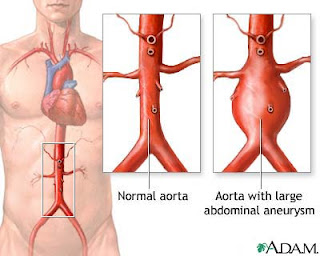
Abdominal Aortic Aneurysm
- Usually in the infrarenal segment of the aorta (80%)
- These most commonly occur below the level of the renal artery
- Features of pain:
o Rapid expansion or rupture will cause epigastric pain radiating to the back. Pain may also be present in the groin, iliac fossae and testicles.
o Can be a constant or intermittent pain
o Be careful not to dismiss it as renal colic!
o
Thoracic Aortic Aneurysm
- Asymmetrical brachial/radial/carotid pulses if the dissection involves the aortic arch. Variable pattern depending on where the dissection is.
o BP may be different in each arm – under similar circumstances to the above.
Pathology of aneurysm
An aneurysm is a permanent dilation of the vessel wall. The fact that it is permanent implies that the vessel wall itself is altered in some way.
Atheromatous degeneration is the most common cause of true aneurysm. Thus the risk factors are the same as for CHD:
- Smoking
- Family history
- Diabetes
- Hypertension
- Age
- Hyperlipidaemia
Most probably pathology
- There is ischaemia of the aortic media where there is an atherosclerotic plaque. This is as a result of release of macrophage enzymes (released when macrophages become activatived) that break down the elastic fibres (collagen and elastin)
o There is evidence that various genetic varients of collagen are more susceptible, and this probably accounts for the familial aspect of aneurysms.
- Where this ischaemia occurs there is loss of the normal elastic nature of the media, allowing it to expand.
Marfan’s syndrome
A connective tissue disorder, and is sometimes (but not always) inherited in an autosomal dominant manner. It iscaused by mutations of the fibrinin gene on chromosome 15. It is very common, and is thought to affect about 1 in 5000 individuals, 25% of which will be the result of a new mutation.
Males and females are equally affected.
Fibrillin-1 gene mutations can be seen in 80% of cases, and aid diagnosis. Testing for this can also be used to screen other family members in known cases.
The most common clinical features are in the musculoskeletal system:
- Arachnodactyly – abnormally long and thin fingers in comparison to the palm. Fingers may also be bent backwards at the MCP to 180’ in some cases.
- Joint hypermobility
- Scoliosis – lateral curvature of the spine
- Chest deformity
- High arched palate
- Dislocation of lens in the eye
- Patients are usually tall and thin, with long limbs
These are generally mild, features, but the disease can also have serious complications, including:
- Heart valve defects
- Pre-disposes to aneurysms
o There is weakening of the media layer of the aorta, leading to dilation. In these cases, the dilatation typically occurs in the ascending aorta. There may also be valve defects (e.g. aortic regurg) which complicate the issue.
o In Marfan’s Syndrome, the root of the aorta is typically affected
- Lung disorders
- Dura disorders
It is thought that as well as the fibrin defects, there are also problems in TGF-β (transforming-growth-factor-β). This is thought to accumulate in heart valve and blood vessels, and alter their underlying structure, leading to the complications mentioned above.
Treatment of Marfan’s
- β-blocker therapy – has been proven to reduce the rate/risk of dilatation of the aorta
- Monitoring of aortic dilatation –via X-Ray, Echo, MRI or CT can be useful in patients with known Marfan’s. Usually pataients are followed up with yearly echo to assess the size of the aorta. In some cases, elective replacement of the ascending aorta may be recommended, to prevent dissection but has a mortality of 5-10%.
- Avoidance of endurance sports / activities
- In pregnancy – as both pregnancy and Marfan’s are risk factors for AA, then during pregnancy, the aorta is closely monitored by 6 weekly echo’s.
o If the aortic root >4cm, ceasarian section should be considered
o β – blocker therapy is safe to continue during pregnancy
Prognosis
- generally good, although less than the general population.
- Surgical interventions have increased the average life-expectancy by 13 years
Notes by Tom Leach

EmoticonEmoticon