Showing posts with label Pathology. Show all posts

Pathology: Cellular Reaction to Injury

0
Notes to Pathology: Cellular Reaction to Injury

I. ADAPTATION TO ENVIRONMENTAL STRESS
A. Hypertrophy
·         Hypertrophy is an increase in the size of an organ or tissue due to an increase in the size of cells.
·         Other characteristics include an increase in protein synthesis and an increase in the size or number of intracellular organelles.
·         A cellular adaptation to increased workload results in hypertrophy, as exemplified by the increase in skeletal muscle mass associated with exercise and the enlargement of the left ventricle in hypertensive heart disease.

B. Hyperplasia
·         Hyperplasia is an increase in the size of an organ or tissue caused by an increase in the number of cells.
·         It is exemplified by glandular proliferation in the breast during pregnancy.
·         In some cases, hyperplasia occurs together with hypertrophy. During pregnancy, uterine enlargement is caused by both hypertrophy and hyperplasia of the smooth muscle cells in the uterus.

C. Aplasia
·         Aplasia is a failure of cell production.
·         During fetal development, aplasia results in agenesis, or absence of an organ due to failure of production.
·         Later in life, it can be caused by permanent loss of precursor cells in proliferative tissues, such as the bone marrow.

D. Hypoplasia
·         Hypoplasia is a decrease in cell production that is less extreme than in aplasia.
·         It is seen in the partial lack of growth and maturation of gonadal structures in Turner syndrome and Klinefelter syndrome.

E. Atrophy
·         Atrophy is a decrease in the size of an organ or tissue and results from a decrease in the mass of preexisting cells.
·         Most often, causal factors are disuse, nutritional or oxygen deprivation, diminished endocrine stimulation, aging, and denervation (lack of nerve stimulation in peripheral muscles caused by injury to motor nerves).
·         Characteristic features often include the presence of autophagic granules, which are intracytoplasmic vacuoles containing debris from degraded organelles.
·         In some instances, atrophy is thought to be mediated in part by the ubiquitin-proteosome pathway of protein degradation. In this pathway, ubiquitin-linked proteins are degraded within the proteosome, a large cytoplasmic protein complex.

F. Metaplasia is the replacement of one differentiated tissue by another
  • Squamous metaplasia
    • Squamous metaplasia is exemplified by the replacement of columnar epithelium at the squamocolumnar junction of the cervix by squamous epithelium.
    • It can also occur in the respiratory epithelium of the bronchus, in the endometrium, and in the pancreatic ducts.
    • Associated conditions include chronic irritation (e.g., squamous metaplasia of the bronchi with long-term use of tobacco) and vitamin A deficiency.
    • This process is often reversible.
  • Osseous metaplasia
    • Osseous metaplasia is the formation of new bone at sites of tissue injury.
    • Cartilaginous metaplasia may also occur.
  • Myeloid metaplasia (extramedullary hematopoiesis) is proliferation of hematopoietic tissue at sites other than the bone marrow, such as the liver or spleen.
II. HYPOXIC CELL INJURY
A. Causes. Hypoxic cell injury results from cellular anoxia or hypoxia, which in turn results from various mechanisms, including:
  • Ischemia (obstruction of arterial blood flow), which is the most common cause
  • Anemia, which is a reduction in the number of oxygen-carrying red blood cells
  • Carbon monoxide poisoning, which results in diminution in the oxygen-carrying capacity of red blood cells by chemical alteration of hemoglobin
  • Decreased perfusion of tissues by oxygen-carrying blood, which occurs in cardiac failure, hypotension, and shock
  • Poor oxygenation of blood secondary to pulmonary disease
B. Early stage. Hypoxic cell injury first affects the mitochondria, with resultant decreased oxidative phosphorylation and adenosine triphosphate (ATP) synthesis. Consequences of decreased ATP availability include:
  • Failure of the cell membrane pump (ouabain-sensitive Na+-K+-ATPase) results in increased intracellular Na+ and water and decreased intracellular K+. This process causes cellular swelling and swelling of organelles.
    • Cellular swelling, or hydropic change, is characterized by the presence of large vacuoles in the cytoplasm.
    • Swelling of the endoplasmic reticulum is one of the first ultrastructural changes evident in reversible injury.
    • Swelling of the mitochondria progresses from reversible, low-amplitude swelling to irreversible, high-amplitude swelling, which is characterized by marked dilation of the inner mitochondrial space.
  • Disaggregation of ribosomes leads to failure of protein synthesis. Ribosomal disaggregation is also promoted by membrane damage.
  • Stimulation of phosphofructokinase activity results in increased glycolysis, accumulation of lactate, and decreased intracellular pH. Acidification causes reversible clumping of nuclear chromatin.
C. Late stage
  • Hypoxic cell injury eventually results in membrane damage to plasma and to lysosomal and other organelle membranes, with loss of membrane phospholipids.
  • Reversible morphologic signs of damage include the formation of:
    • Myelin figures, whorl-like structures probably originating from damaged membranes
    • Cell blebs, a cell surface deformity most likely caused by disorderly function of the cellular cytoskeleton
D. Cell death. Finally, cell death is caused by severe or prolonged injury.
  • The point of no return is marked by irreversible damage to cell membranes, leading to massive calcium influx, extensive calcification of the mitochondria, and cell death.
  • Intracellular enzymes and various other proteins are released from necrotic cells into the circulation as a consequence of the loss of integrity of cell membranes. This phenomenon is the basis of a number of useful laboratory determinations as indicators of necrosis.
    • Myocardial enzymes in serum.
      • Enzymes that have been useful in the diagnosis of myocardial infarction include the following:
        • Aspartate aminotransferase (AST, previously known as SGOT)
        • Lactate dehydrogenase (LDH)
        • Creatine kinase (CK, also known as CPK)
      • These markers of myocardial necrosis vary in specificity for heart damage, as well as in the time period after the necrotic event in which elevations in the serum appear and persist. The delineation of isoenzyme forms of LDH and CK has been a useful adjunct in adding specificity to these measures.
      • The foregoing enzymes are beginning to be replaced by other myocardial proteins in serum as indicators of myocardial necrosis. Important examples include the troponins (troponin I [TnI] and troponin T [TnT]) and myoglobin.
    • Liver enzymes in serum. Enzymes of special interest include the transaminases (AST and alanine aminotransferase [ALT]), alkaline phosphatase, and >-glutamyltransferase (GGT).
  • The vulnerability of cells to hypoxic injury varies with the tissue or cell type. Hypoxic injury becomes irreversible after:
    • 3–5 minutes for neurons. Purkinje cells of the cerebellum and neurons of the hippocampus are more susceptible to hypoxic injury than are other neurons.
    • 1–2 hours for myocardial cells and hepatocytes
    • Many hours for skeletal muscle cells
III. FREE RADICAL INJURY
A. Free radicals
  • These molecules have a single unpaired electron in the outer orbital.
  • Examples include the activated products of oxygen reduction, such as the superoxide (O2-) and the hydroxyl (OH·) radicals.
B. Mechanisms that generate free radicals
  • Normal metabolism
  • Oxygen toxicity, such as in the alveolar damage that can cause adult respiratory distress syndrome or as in retrolental fibroplasia (retinopathy of prematurity), an ocular disorder of premature infants that leads to blindness
  • Ionizing radiation
  • Ultraviolet light
  • Drugs and chemicals, many of which promote both proliferation of the smooth endoplasmic reticulum (SER) and induction of the P-450 system of mixed function oxidases of the SER. Proliferation and hypertrophy of the SER of the hepatocyte are classic ultrastructural markers of barbiturate intoxication.
  • Reperfusion after ischemic injury
C. Mechanisms that degrade free radicals
  • Intracellular enzymes, such as glutathione peroxidase, catalase, or superoxide dismutase
  • Exogenous and endogenous antioxidants, such as vitamin A, vitamin C, vitamin E, cysteine, glutathione, selenium, ceruloplasmin, or transferrin
  • Spontaneous decay
IV. CHEMICAL CELL INJURY
Chemical cell injury is illustrated by the model of liver cell membrane damage induced by carbon tetrachloride (CCl4).
  • In this model, CCl4 is processed by the P-450 system of mixed function oxidases within the SER, producing the highly reactive free radical CCl3·.
  • CCl diffuses throughout the cell, initiating lipid peroxidation of intracellular membranes. Widespread injury results, including:
    • Disaggregation of ribosomes, resulting in decreased protein synthesis. Failure of the cell to synthesize the apoprotein moiety of lipoproteins causes an accumulation of intracellular lipids (fatty change).
    • Plasma membrane damage, caused by products of lipid peroxidation in the smooth endoplasmic reticulum, resulting in cellular swelling and massive influx of calcium, with resultant mitochondrial damage, denaturation of cell proteins, and cell death
A. General considerations
  • Necrosis is one of two contrasting morphologic patterns of tissue death. The other is apoptosis (see VI).
  • Necrosis is the sum of the degradative and inflammatory reactions occurring after tissue death caused by injury (e.g., hypoxia, exposure to toxic chemicals); it occurs within living organisms. In pathologic specimens, fixed cells with well-preserved morphology are dead but not necrotic.
  • Autolysis refers to degradative reactions in cells caused by intracellular enzymes indigenous to the cell. Postmortem autolysis occurs after the death of the entire organism and is not necrosis.
  • Heterolysis refers to cellular degradation by enzymes derived from sources extrinsic to the cell (e.g., bacteria, leukocytes).
B. Types of necrosis
  • Coagulative necrosis
    • Coagulative necrosis results most often from a sudden cutoff of blood supply to an organ (ischemia), particularly the heart and kidney.
    • General preservation of tissue architecture is characteristic in the early stages.
    • Increased cytoplasmic eosinophilia occurs because of protein denaturation and loss of cytoplasmic RNA.
    • Nuclear changes, the morphologic hallmark of irreversible cell injury and necrosis, are characteristic. These include:
      • Pyknosis, chromatin clumping and shrinking with increased basophilia
      • Karyorrhexis, fragmentation of chromatin
      • Karyolysis, fading of chromatin material
      • Disappearance of stainable nuclei
  • Liquefactive necrosis
    • Ischemic injury to the central nervous system (CNS) characteristically results in liquefactive necrosis. After the death of CNS cells, liquefaction is caused by autolysis.
    • Digestion, softening, and liquefaction of tissue are characteristic.
    • Suppurative infections characterized by the formation of pus (liquefied tissue debris and neutrophils) by heterolytic mechanisms involve liquefactive necrosis.
  • Caseous necrosis
    • This type of necrosis occurs as part of granulomatous inflammation and is a manifestation of partial immunity caused by the interaction of T lymphocytes (CD4+, CD8+, and CD4-CD8-), macrophages, and probably cytokines, such as interferon-γ, derived from these cells.
    • Tuberculosis is the leading cause of caseous necrosis.
    • Caseous necrosis combines features of both coagulative necrosis and liquefactive necrosis.
    • On gross examination, caseous necrosis has a cheese-like (caseous) consistency.
    • On histologic examination, caseous necrosis has an amorphous eosinophilic appearance.
  • Gangrenous necrosis
    • This type of necrosis most often affects the lower extremities or bowel and is secondary to vascular occlusion.
    • When complicated by infective heterolysis and consequent liquefactive necrosis, gangrenous necrosis is called wet gangrene.
    • When characterized primarily by coagulative necrosis without liquefaction, gangrenous necrosis is called dry gangrene.
  • Fibrinoid necrosis
    • This deposition of fibrin-like proteinaceous material in the arterial walls appears smudgy and acidophilic.
    • Fibrinoid necrosis is often associated with immune-mediated vascular damage.
  • Fat necrosis occurs in two forms.
    • Traumatic fat necrosis, which occurs after a severe injury to tissue with high fat content, such as the breast
    • Enzymatic fat necrosis, which is a complication of acute hemorrhagic pancreatitis, a severe inflammatory disorder of the pancreas
      • Proteolytic and lipolytic pancreatic enzymes diffuse into inflamed tissue and literally digest the parenchyma.
      • Fatty acids liberated by the digestion of fat form calcium salts (saponification, or soap formation).
      • Vessels are eroded, with resultant hemorrhage.
A. General considerations
  • Apoptosis is a second morphologic pattern of tissue death. (The other is necrosis; see V.) It is often referred to as programmed cell death.
  • This is an important mechanism for the removal of cells. An example is apoptotic removal of cells with irreparable DNA damage (from free radicals, viruses, cytotoxic immune mechanisms), protecting against neoplastic transformation
  • In addition, apoptosis is an important mechanism for physiologic cell removal during embryogenesis and in programmed cell cycling (e.g., endometrial cells during menstruation).
  • This involutional process is similar to the physiologic loss of leaves from a tree; apoptosis is a Greek term for "falling away from."
B. Morphologic features
  • A tendency to involve single isolated cells or small clusters of cells within a tissue
  • Progression through a series of changes marked by a lack of inflammatory response
    • Blebbing of plasma membrane, cytoplasmic shrinkage, chromatin condensation
    • Budding of cell and separation of apoptotic bodies (membrane-bound segments)
    • Phagocytosis of apoptotic bodies
  • Involution and shrinkage of affected cells and cell fragments, resulting in small round eosinophilic masses often containing chromatin remnants, exemplified by Councilman bodies in viral hepatitis
C. Biochemical events
  • Diverse injurious stimuli (e.g., free radicals, radiation, toxic substances, withdrawal of growth factors or hormones) trigger a variety of stimuli, including cell surface receptors such as FAS, mitochondrial response to stress, and cytotoxic T cells.
  • The extrinsic pathway of initiation is mediated by cell surface receptors exemplified by FAS, a member of the tumor necrosis factor receptor family of proteins. This pathway is initiated by signaling by molecules such as the FAS ligand, which in turn signals a series of events that involve activation of caspases. Caspases are aspartate-specific cysteine proteases that have been referred to as "major executioners" or "molecular guillotines." The death signals are conveyed in a proteolytic cascade, through activation of a chain of caspases and other targets. The initial activating caspases are caspase-8 and caspase-9, and the terminal caspases (executioners) include caspase-3 and caspase-6 (among other proteases).
  • The intrinsic, or mitochondrial, pathway, which is initiated by the loss of stimulation by growth factors and other adverse stimuli, results in the inactivation and loss of bcl-2 and other antiapoptotic proteins from the inner mitochondrial membrane. This loss results in increased mitochondrial permeability, the release of cytochrome c, and the stimulation of proapoptotic proteins such as bax and bak. Cytochrome c interacts with Apaf-1 causing self-cleavage and activation of caspase-9. Downstream caspases are activated by upstream proteases and act themselves to cleave cellular targets.
  • Cytotoxic T-cell activation is characterized by direct activation of caspases by granzyme B, a cytotoxic T-cell protease that perhaps directly activates the caspase cascade. The entry of granzyme B into target cells is mediated by perforin, a cytotoxic T-cell protein.
  • Degradation of DNA by endonucleases into nucleosomal chromatin fragments that are multiples of 180–200 base pairs results in the typical "laddering" appearance of DNA on electrophoresis. This phenomenon is characteristic of, but not entirely specific for, apoptosis.
  • Activation of transglutaminases crosslinks apoptotic cytoplasmic proteins.
  • The caspases consist of a group of aspartic acid-specific cysteine proteases that are activated during apoptosis.
  • Newer methods such as the TUNEL assay (Terminal Transferase dUTP Nick End Labeling) are ways to quantitate cleaving of nucleosomes and, thus, apoptosis. Similarly, caspase assays are coming into use as apoptotic markers. Surely more will follow.
D. Regulation of apoptosis is mediated by a number of genes and their products. Important genes include bcl-2 (gene product inhibits apoptosis), bax (gene product facilitates apoptosis), and p53 (gene product decreases transcription of bcl-2 and increases transcription of bax, thus facilitating apoptosis).
E. Additionally, complex signaling pathways involving multiple genes and gene products are the subject of vigorous scientific investigation. Since many pathologic processes are related to either stimulation or inhibition of apoptosis (e.g., many forms of cancer), this area of inquiry promises to yield major understanding that will surely lead to important therapeutic applications.


Hereditary hemochromatosis
Hereditary hemochromatosis. Prussian blue staining marks the intraparenchymal deposition of hemosiderin.

VII. REVERSIBLE CELLULAR CHANGES AND ACCUMULATIONS
A. Fatty change (fatty metamorphosis, steatosis)
  • General considerations
    • Fatty change is characterized by the accumulation of intracellular parenchymal triglycerides and is observed most frequently in the liver, heart, and kidney. For example, in the liver, fatty change may be secondary to alcoholism, diabetes mellitus, malnutrition, obesity, or poisonings.
  • Imbalance among the uptake, utilization, and secretion of fat is the cause of fatty change, and this can result from any of the following mechanisms:
    • Increased transport of triglycerides or fatty acids to affected cells
    • Decreased mobilization of fat from cells, most often mediated by decreased production of apoproteins required for fat transport. Fatty change is thus linked to the disaggregation of ribosomes and consequent decreased protein synthesis caused by failure of ATP production in CCl4-injured cells.
    • Decreased use of fat by cells
    • Overproduction of fat in cells
B. Hyaline change
  • This term denotes a characteristic (homogeneous, glassy, eosinophilic) appearance in hematoxylin and eosin sections.
  • It is caused most often by nonspecific accumulations of proteinaceous material.
C. Accumulations of exogenous pigments
  • Pulmonary accumulations of carbon (anthracotic pigment), silica, and iron dust
  • Plumbism (lead poisoning)
  • Argyria (silver poisoning), which may cause a permanent gray discoloration of the skin and conjunctivae
D. Accumulations of endogenous pigments
  • Melanin
    • This pigment is formed from tyrosine by the action of tyrosinase, synthesized in melanosomes of melanocytes within the epidermis, and transferred by melanocytes to adjacent clusters of keratinocytes and also to macrophages (melanophores) in the subjacent dermis.
    • Increased melanin pigmentation is associated with suntanning and with a wide variety of disease conditions.
    • Decreased melanin pigmentation is observed in albinism and vitiligo.
  • Bilirubin
    • This pigment is a catabolic product of the heme moiety of hemoglobin and, to a minor extent, myoglobin.
    • In various pathologic conditions, bilirubin accumulates and stains the blood, sclerae, mucosae, and internal organs, producing a yellowish discoloration called jaundice.
      • Hemolytic jaundice, which is associated with the destruction of red cells.
      • Hepatocellular jaundice, which is associated with parenchymal liver damage, and obstructive jaundice, which is associated with intra- or extrahepatic obstruction of the biliary tract.
  • Hemosiderin
    • This iron-containing pigment consists of aggregates of ferritin. It appears in tissues as golden brown amorphous aggregates and can be positively identified by its staining reaction (blue color) with Prussian blue dye. It exists normally in small amounts as physiologic iron stores within tissue macrophages of the bone marrow, liver, and spleen.
    • It accumulates pathologically in tissues in excess amounts (sometimes massive).
      • Hemosiderosis is defined by accumulation of hemosiderin, primarily within tissue macrophages, without associated tissue or organ damage.
      • Hemochromatosis is more extensive accumulation of hemosiderin, often within parenchymal cells, with accompanying tissue damage, scarring, and organ dysfunction. This condition occurs in both hereditary (primary) and secondary forms.
        • Hereditary hemochromatosis is most often caused by a mutation in the Hfe gene on chromosome 6.
          • Hemosiderin deposition and organ damage in the liver, pancreas, myocardium, and multiple endocrine glands is characteristic, as well as melanin deposition in the skin.
          • This results in the triad of micronodular cirrhosis, diabetes mellitus,skin pigmentation. This set of findings is referred to as "bronze diabetes." Laboratory abnormalities of note include marked elevation of the serum transferrin saturation because of the combination of increased serum iron and decreased total iron-binding capacity (TIBC). and
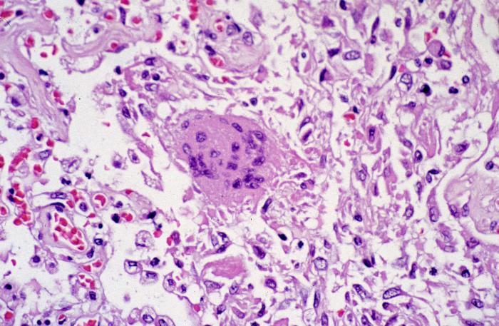
        • Secondary hemochromatosis is most often caused by multiple blood transfusions administered to subjects with hereditary hemolytic anemias such as β-thalassemia major (Figure 1-4).
  • Lipofuscin
    • This yellowish, fat-soluble pigment is an end product of membrane lipid peroxidation.
    • It is sometimes referred to as "wear-and-tear" pigment.
    • It commonly accumulates in elderly patients, in whom the pigment is found most often within hepatocytes and at the poles of nuclei of myocardial cells. The combination of lipofuscin accumulation and atrophy of organs is referred to as brown atrophy.
E. Pathologic calcifications
  • Metastatic calcification
    • The cause of metastatic calcification is hypercalcemia.
    • Hypercalcemia most often results from any of the following causes:
      • Hyperparathyroidism
      • Osteolytic tumors with resultant mobilization of calcium and phosphorus
      • Hypervitaminosis D
      • Excess calcium intake, such as in the milk-alkali syndrome (nephrocalcinosis and renal stones caused by milk and antacid self-therapy)
  • Dystrophic calcification
    • Dystrophic calcification is defined as calcification in previously damaged tissue, such as areas of old trauma, tuberculosis lesions, scarred heart valves, and atherosclerotic lesions.
    • The cause is not hypercalcemia; typically, the serum calcium concentration is normal (Figure 1-5).
VIII. DISORDERS CHARACTERIZED BY ABNORMALITIES OF PROTEIN FOLDING
A. These disorders involve failure of protein structural stabilization or degradation by specialized proteins known as chaperones. Important chaperones include heat shock proteins induced by stress, one of which is ubiquitin, which marks abnormal proteins for degradation.
B. Two known pathogenetic mechanisms include:
  • Abnormal protein aggregation, which is characteristic of amyloidosis; a number of neurodegenerative diseases, such as Alzheimer disease, Huntington disease, and Parkinson disease; and perhaps prion diseases, such as "mad cow" disease
  • Abnormal protein transport and secretion, which is characteristic of cystic fibrosis and α1-antitrypsin deficiency


Goljan - Audio Transcript

0
goljan audio transcript
Goljan - Audio Transcript
This is 125 pages someone wrote after the audio files of Dr Goljan pathology lectures for step1 (thanks for him) It's really helpful for those who find difficulty to follow his rapid sequence of facts in some files (including me) it's really helpful for revision after the audio files and it's ordered as the same order of audio files. It is good for USMLE step 1.


Click below:

DOWNLOAD 

Outlines of Pathology - eBook

0

Pathology: Outlines of Pathology - eBook

This is a good review of patholgy by:

outlines of pathology

John H. Sinard, MD, PhD


Over the years, a number of pathology residents have commented that they found this book to be a valuable resource when learning pathology and studying for the boards. In the interests of continuing to keep this work available to Pathologists and Pathology Residents who might still find it useful.


    DOWNLOAD


    or

    DOWNLOAD


    Pathology High Yield USMLE Step1 - Goljan

    0
    This is the 20 pages long review of pathology, suitable for USMLE Step1.

    Click Below to download:


    DOWNLOAD

    GOLJAN - Systemic Pathology

    0
    Dr. Edward Goljan - Systemic Pathology notes - DOWNLOAD LINK BELOW!
    Pages: 277

    Goljan Systemic Pathology:

    Scribd
    Or
    DOWNLOAD
    Pathology: Queen Mary, UOL Notes

    Pathology: Queen Mary, UOL Notes

    0
     Pathology: Queen Mary, UOL Notes
    The text is presented as Word files. Your computer will open them when you click on them, or you can download them. There are various disadvantages about these notes.
    1. They are not finished
    2. There is no index
    3. The pictures have not all been done! Those which have been done are in Coreldraw. They comprise many of the immunology pictures, and one microbiology picture. They will be given out as a booklet.
    There are some advantages.
    1. They are free.
    2. They are being continually updated.
    3. You can come and argue with the author (me).
    Good luck, and best wishes, Nigel Yeatman, 2004
    Infectious agents
    The defence system
    Subsection 1:Introduction
    Subsection 2: innate components of the immune system.
    Subsection 3: B lymphocytes and antibodies
    Subsection 4: Antibody genes and B-lymphocyte development
    Subsection 5: Antigen presenting cells and T-lymphocytes
    Subsection 6: CD4 T-cells
    Subsection 7: CD8 T-cells
    Subsection 8: HLA molecules and their genes
    Subsection 9: T-lymphocyte receptor genes, T-lymphocyte development, and the thymus
    Subsection 10: Other populations of T-cells, and Null cells.
    Subsection 11: lymphoid tissues, and the circulation of cells of the immune system.
    Subsection 12: Tjhe innate immune system revisited: the effect of the immune response.
    Subsection 13: Acute inflammation
    Subsection 14: Wound healing anfd repair
    Subsection 15: hypersensitivity and autoimmunity.
    Subsection 16: Chronic inflammation.
    Subsection 17: Neoplasia and the immune system.
    Subsection 18: Increased susceptibility to infection.
    Subsection 19: immunisation, ande the upregulation of immunity.
    Subsection 20: Transplantation.
    Subsection 21: Laboratory tests in immunology.
    Subsection 22: Anti-inflammatory and immunosuppressive drugs.

    Neoplasia
    Pathology: General Notes

    Pathology: General Notes

    0

    Impetigo

    Highly contagious bacterial infection of epidermis

    Infectious agent: Staph aureus and Group A Strep

    Common in children

    Signs and symptoms:

    Itchy, fluid filled blisters

    Erosions with honey-colored crusts (Fig. 1.12)

    Usually forms around mouth, nose or ears; can be elsewhere

    Treatment: antibiotics

    Prevention

    Keep open lesions clean and covered when possible

    Massage?

    Systemically contraindicated until the infection has been eradicated

    Folliculitis and Furuncles

    Bacterial infection of a hair follicle and perifollicular dermis

    Infectious agent: Staphylococcus aureus

    Common locations: shaved regions (beard, neck, legs, axilla) or occlusion (axilla, below breasts, buttocks)

    If follicular infection extends into dermis, an acute local inflammatory process with tissue destruction results:

    Furuncle (boil): 1-2cm

    Carbuncle: coalescing furuncles that may develop subQ abscess

    Pilonidal cysts: chronic abscess of follicle near sacrum

    Hidradenitis suppurativa: infection/inflammation of apocrine glands in axilla, anogenital region, breasts

    related to cystic acne; forms double open comedomes

    Signs and symptoms:

    Folliculitis: follicular red bump or pustule

    Furuncle: red, hot, fluctuant, painful nodule

    May develop central necrotic plug or pustule

    Central pustule may rupture and drain pus

    Treatment:

    Hot compresses

    Incision and drainage

    Antibiotics

    Massage?

    At least locally contraindicated

    Isolate sheets

    Watch for signs of systemic infection


    Cellulitis and Erysipelas

    Acute, rapidly spreading bacterial infection of the dermis and subcutaneous tissue (Fig. 1.4)

    Infectious agent:
    Erysipelas: Group A Streptococcus (GAS), H. influenza B in peds (facial)
    Cellulitis: Staph aureus, GAS

    Requires a portal of entry: interdigital tinea pedis, bee sting!

    Signs and symptoms:

    Erysipelas: red, raised, hard, hot, painful area with distinct margin

    esp face (rhinitis, conjunctivitis, OM) and lower legs

    Cellulitis: red, flat, hard, hot, painful area with indistinct margin

    Lymphatic spread (lymphangitis): red streaks proximal to site

    Systemic symptoms: fever, chills, malaise

    Complications: septicemia (potentially fatal)

    Treatment: Antibiotics

    Massage? Systemically contraindicated until infection resolves


    Fungal Infections (Not discussing candidiasis or “yeast infections”)

    Other names:

    Dermatophytosis

    Mycosis

    Ringworm

    Lesions are called tinea

    Causative agents are fungi:

    Tricophyton, Epidermophyton, Microsporum

    Fungi like warm, moist, dark places

    Infection is spread by direct contact (skin to skin or fomite to skin)

    Signs and symptoms:

    Several days’ gestation before symptoms develop; communicable during this time

    Tinea corporis (body ringworm) (Fig. 1.6)

    Itchy, scaled, raised, reddish ring

    Central clearing (heals from center out)

    Spreads with scratching

    Tinea capitis (head ringworm) (Fig. 1.7)

    Itchy, flaking skin; looks like dandruff

    Can cause permanent hair loss

    Tinea pedis (athlete’s foot) (Fig. 1.8)

    Often starts between 3rd and 4th toes

    Weepy blisters; cracked and peeling skin; itching and burning

    Moccasin type: heels, soles, lateral borders of feet; like T. corporis – red with distinct, raised, scaly margin

    Risk of secondary infection

    Difficult to get rid of (treat shoes too)

    Tinea cruris (jock itch) (Fig. 1.9)

    Groin, buttocks, thighs; NOT penis or scrotum

    Itchy, red lesion with distinct margin

    Less contagious than other forms

    T. manus: hands; may be connected to athlete’s foot

    T. barbae: beard; looks like folliculitis

    T. unguium: nails; also known as onychomycosis

    T. versicolor = Pityriasis versicolor:

    Pityrosporum ovale inhibits melanin synthesis

    Typically pales when surrounding skin tans

    Neck, trunk, upper arms

    Treatment:

    Topical or oral anti-fungals

    Sodium selenite shampoo

    Massage?

    Recognize it; avoid it. Be careful about athlete’s foot

    Local contraindication or caution, if well-contained and covered


    Herpes Simplex

    Viral infection leading to painful blisters on the skin

    Oral, genital, other

    HSV-1 v. HSV-2: no longer a significant issue

    Primary Outbreak: days – 3 weeks after exposure

    May be unnoticeable; may be extremely severe with systemic symptoms

    Recurrent herpes: virus is never expelled;

    Virus goes dormant in dorsal root ganglia

    Reactivation of virus triggers another outbreak

    Incidence: Who knows?

    Oral herpes: 80%?

    Genital herpes: 60%?

    Communicability:

    Virus is shed in mucus and respiratory secretions

    Lesion not required for shedding

    Can spread between outbreaks and during prodromic stage

    Virus is stable on surfaces outside a host for hours

    Can spread from one part of the body to another (eye!)

    Vertical spread: mother to vaginally-delivered infant

    Types of herpes:

    Oral: “cold sore” or “fever blister” (Fig. 1.10)

    Genital

    Whitlow: paronychial (Fig. 1.11)

    Gladiatorum: wrestlers

    Triggers:

    Physical or emotional stress

    Hormonal changes

    Sunlight

    High arginine foods (chocolate, nuts)

    Signs and symptoms:

    Tingling, itching, burning during prodromic stage

    Painful blisters on a red base

    Scabbing after acute phase (less contagious)

    Genital herpes: maybe systemic sx and urethritis

    Complications:

    Secondary infection

    Increased risk of spreading HIV

    Increased risk of cervical cancer (HPV)

    Encephalitis/meningitis

    Treatment:

    Antivirals to shorten episode (prevention in frequent cases)

    Massage?

    Systemic Contraindication for systemic symptoms

    Local contraindication during outbreaks, prodromic stage

    Isolate linens and treat accordingly

    Be careful of spreading from therapist to client!


    Warts

    Viral infection of keratinocytes

    Causative agent: human papilloma virus (HPV)

    Spread by skin-to-skin contact

    Virus grows slowly, usually self-limiting

    Causes epidermal hyperplasia – a benign neoplasm

    Types of warts:

    Verruca vulgaris: cauliflower type

    On sites of trauma: fingers, hands, knees

    Verruca plana: flat warts (flat, light brown)

    On dorsum of hand, face, shins

    Verruca plantaris: plantar wart

    On sole of foot or palm of hand

    Genital warts: tag-like growths on penis, vulva, anust, etc.

    NOT associated with cervical cancer

    Different strains of HPV are are spread with sexual contact and linked with cervical dysplasia and cervical cancer

    Signs and symptoms:

    Hard, crusty pile-up of keratin (Fig. 1.19)

    Can resemble callus on the feet; important to distinguish between them (Fig. 1.20)

    Treatment:

    Abrade and apply salicylic acid (compound W), duct tape, raw potato…

    Liquid nitrogen, lasers

    Immune support

    Warts are suggestible–many folk remedies

    Massage?

    Local contraindication (virus is in shedding skin)


    Molluscum Contagiosum

    NOT a type of wart

    Infectious agent: Poxvirus

    Self-limiting epidermal infection (6 months)

    Common in children

    Can be an STD

    Do NOT abrade: this will spread lesions


    Lice and Mites

    Mites or “Scabies”: Sarcoptes scabiei

    Burrow under the skin; fecal waste is allergen

    Spread through intimate contact

    Signs and symptoms of mites:

    Scabies: reddish or grayish tracks on the skin (these are the burrows) (Fig. 1.14)

    Locations: finger webs, ankles, anywhere!

    Unrelenting itchiness, worse at night

    Treatment:

    Pesticidal soap – for body, linens, clothes

    For patient and people in close contact


    Head lice: Pediculus humanus capitis

    Live in head hair; suck blood from scalp

    Spread through soft surfaces (clothing, hats, upholstery, etc.)

    Symptoms of head lice:

    Nits (brown or white) (Fig. 1.16)

    Itching

    Secondary dermatitis

    Head lice treatment:

    Pesticidal shampoo

    Nit comb, nit-picking


    Body Lice: Pediculus humanus humans

    Live in clothing, not on hosts

    Eradicated with good hygiene, tea tree oil soap


    Pubic Lice or “Crabs”: Pthirus pubis

    Infest pubic hair and any other coarse body hair (Fig. 1.18)

    Spread through intimate contact

    Itching, may be worse at night

    Pesticidal shampoo (like head lice)

    Massage for clients with lice or mites?

    Systemically contraindicated until the infestation is over


    Acne Vulgaris

    Inflammation of pilosebaceous unit, usually on face, chest, or back (Fig. 1.21)

    Sterile inflammatory response to excess sebum and bacterial fatty acids causes hyperkeratinization → keratin plug in os of follicle = comedone

    If comedone ruptures and leak into dermis, immune response → pustule, nodule, cyst with inflammation

    Occurs in up to 80% of the population

    Contributing factors:

    Hormonal imbalances

    Testosterone production

    Liver congestion

    Bacterial activity: Propionibacterium acnes

    Stress

    Signs and symptoms (types of lesions):

    Open comedones: blackhead (oxidized keratin)

    Closed comedones: whitehead (keratin plug)

    Pimples: pustule

    Cysts: large comedomes due to repeated rupture and recapsulation

    Treatment:

    Hygiene

    Liver support

    Rx: Antibiotics, Retinoids, OCPs

    Massage?

    Local contraindication – may aggravate/spread lesions

    Don’t use alcohol after a session (???)

    Consider water-based lotion

    Rosacea (not often called Acne rosacea)

    Idiopathic chronic skin condition

    Affects the skin mostly around the nose and cheeks

    Triggers include:

    Sunlight, wind, extreme temperatures

    Food and drinks

    Stress

    Signs and symptoms: 4 Stages:

    Pre-rosacea: flushing, no itch

    Vascular rosacea: telangiectasias develop; the nose, eyes, and eyelids may be involved

    Inflammatory rosacea: papules and pustules develop, but not the same causative agent as acne vulgaris

    Rhinophyma: skin is permanently red, thickened, bumpy

    Treatment:

    Palliative interventions to reduce appearance of lesions

    Antibiotics

    Avoidance of triggers

    Massage?

    No specific cautions unless work on the face increases symptoms; other work is safe


    Dermatitis/Eczema

    Dermatitis = skin inflammation

    Eczema = to boil over

    Vague enough?

    Not from an infectious source

    Epidermal eruptions with intercellular edema

    Acute: Itchy vesicles or juicy papules (edema)

    Chronic: Itchy lichenification (hyperkeratosis)

    Related to hypersensitivity reactions

    Eczema

    Eczema: overreactive systemic immune response to an internal allergen that manifests in skin

    Incidence: affects 1:7 babies and children

    “Grow out of it”: only 3% adults

    Cause: Type I Hypersensitivity Reaction

    B-cells release IgE antibodies against specific foreign protein (antigen)

    Activated mast cells release histamine and other cytokines

    Massive, rapid inflammatory response

    Also associated with:

    Genetics: family history of atopic disease (eczema, allergic rhinitis/sinusitis, asthma)

    Fatty acid deficiency

    Monocyte malfunction, high levels IgE, excessive histamine

    Food allergy

    May be aggravated by harsh detergents and chemicals, irritating fabrics, climate, stress, etc.

    Eczema signs and symptoms (types of eczema):

    Atopic Dermatitis = classic eczema

    Most common

    Infants: head, diaper area

    Children/Adults: face, neck, wrists, dorsum of hands/feet, joint flexures

    Itchy red bumps/blisters

    Patches of itchy, red, flaky, dry skin (Fig. 1.22)

    Can be weepy or so dry it cracks or lichenified

    Dyshidrotic Eczema

    Associated with atopic disease

    Itchy, weeping blisters on palms, soles, or sides of digits (Fig. 1.23)

    Nummular Eczema

    Circular/oval red, weeping patches with crusts

    Commonly trunk, legs and buttocks (Fig. 1.24)

    Seborrheic Dermatitis = cradle cap

    Chronic, superficial inflammatory response to Pityrosporum ovale in hairy regions with active sebaceous glands

    Red patches with yellowish greasy scaling

    Scalp, eyebrows, nose, pre-sternum and skin folds


    Contact Dermatitis

    Contact irritant dermatitis:

    Due to substance directly toxic to skin (acids, alkalis, solvents)

    Anyone would have a similar reaction

    Phototoxic reactions: lime juice, celery, carrot greens, drugs

    Contact allergic dermatitis: Type IV Hypersensitivity Reaction

    Allergic reaction at the site of contact 24-48 hrs after exposure

    Common allergens:

    Nickel (watchband, earrings, etc.) (Fig. 1.25)

    Ingredients in personal hygiene products

    Adhesives, latex

    Poison ivy, oak, sumac: oleoresins

    Photoallergic reactions:

    PABA (sunblock)

    St John’s Wort

    Signs and symptoms of contact dermatitis:

    Range from mildly irritated to acutely red, swollen itchy

    Other types of dermatitis:

    Neurodermatitis = lichen simplex chronicus

    Confluence of small eczematous eruptions that eventually lichenify

    Due to chronic scratching!

    Stasis dermatitis

    Related to poor circulation: venous insufficiency or stasis (thrombophlebitis, DM, CHF)

    Pitting edema; dusky red discoloration; thickened, scaly, weepy, itchy

    May ulcerate

    Complications of eczema/contact dermatitis:

    Secondary infection

    Itch-scratch cycle

    Treatment:

    Avoid triggers

    Good hydration of skin

    Topical steroids (have some risks) or immunomodulators

    Massage?

    It depends on…

    The severity of the problem

    If the skin is intact

    Local Contraindications:

    Active eczema: open, itchy, inflamed

    Active dyshidrotic eczema

    Active contact dermatitis

    Active neurodermatitis

    Systemic Contraindications:

    All acute dermatitis until inflammation has subsided

    Stasis dermatitis: no circulatory massage


    Hives

    Also called urticaria

    Allergic: Type I Hypersensitivity Reaction activates cutaneous mast cells

    Food allergy

    Bee sting

    Medication

    Stress reactions

    Emotions

    Physical: Sunlight, Heat/sweating, Cold, Pressure

    Signs and symptoms:

    Acute dermal edema

    Wheal: Hot, red, itchy, raised (edematous) areas with central pallor

    Individual wheals that may join together in larger patches

    Dermographism: wheal and flare response to scratching skin

    Angioedema: acute dermal and subQ edema (usually larger area)

    Treatment:

    Antihistamines; steroidal anti-inflammatories

    Massage?

    Systemic Contraindication: acute hives; angioedema (esp if respiratory symptoms)

    Local contraindication: subacute

    Be careful about allergenic oils


    Psoriasis

    Abnormal epidermal proliferation resulting in accumulation of stratum corneum & inflammation

    Epidermis replaced in 2-4 days vs. 30 days

    Flares and remission

    Cause is not well understood

    Genetic: 1/3 cases

    T-cell dysfunction: autoimmune disease?

    Vitamin D deficiency?

    Triggers

    Physical trauma: rubbing, scratching

    Stress

    Food allergies?

    Strep throat (guttate psoriasis)

    Incidence:

    About 5 million US

    Types of psoriasis

    Psoriasis vulgaris: most common form

    Guttate psoriasis: <2%

    Explosion of small circular lesions on trunk

    Typically after strep infection

    Pustular psoriasis: rare

    Small non-infectious pustules on plaques or palms/soles

    Erythroderma: very rare

    Generalized psoriasis with extreme redness and systemic symptoms

    Can be life threatening

    Signs and symptoms:

    Itchy psoriatic plaques

    Raised reddish lesions with silvery scale and sharp margins (Figs. 1.28, 1.29)

    Distribution: scalp, hairline, extensor surface of joints, intergluteal cleft

    Inverse pattern: skin folds (umbilicus, axilla, inframammary, inguinal, integluteal fold) and genitalia (no scale)

    Pitted nails

    Psoriatic arthritis: 5% cases; DIPs

    Complications:

    2° Infection

    Treatment:

    Topical applications

    Oral medications: steroids, retinoids, psoralen, cytotoxic drugs

    Treatments may be applied with UV radiation

    Most treatments are temporarily successful

    Massage?

    Local contraindication acutely

    Watch for signs of cracking or compromised skin


    Moles

    Also called nevi

    Benign neoplasm

    Many melanocytes making melanin in one spot

    Appear between age 6 mos and 35 years

    Signs and symptoms:

    Typically small (under 5 mm)

    One-colored: tan to blue-black

    Round or oval (symmetrical)

    Flat or raised, smooth or warty

    May grow course, dark hairs

    Note: can darken or itch during adolescence and pregnancy

    Risk of changing to melanoma:

    Any change in a mole should be examined by a dermatologist

    Large congenital nevus: 6-12% risk

    Dysplastic nevus (atypical mole): ABCDE

    Asymmetry in shape

    Border is irregular, indistinct, or ill-defined

    Color is mottled or mixed (brown, black, purple0

    Diameter is usually large, greater than the tip of a pencil eraser (6.0 mm)

    Enlargement or increased elevation is one of the most important signs of malignant melanoma

    Treatment:

    Nothing

    Removing them

    Massage?

    No contraindications

    Therapists may be able to see moles that clients cannot

    Skin Cancer

    Cells in the epidermis suffer genetic mutations and begin to replicate uncontrollably

    Most commonly diagnosed form of cancer (1/3)

    40-50% people over 65 years

    Special risk factors:

    Pale skin

    History of severe sunburn; sunny climate, spends time outside

    Immune compromised

    Increasing age

    Toxic exposures

    Red Flags

    Moles that change: ABCDE

    Any sore that doesn’t heal

    Massage for skin cancer? Depends on the type of cancer, and the types of treatments

    AK, BCC are safest for massage; locally avoid lesions and/or recent incision sites

    SCC, malignant melanoma may require adjustments based on treatment options; consult with the oncologist for more information

    Actinic Keratosis

    Actinic Keratosis (AK): a precancerous neoplasm (Figs. 1.30, 1.31)

    On lips: actinic cheilitis

    Inside mouth: leukoplakia

    Bowen’s disease: squamous cell carcinoma in situ

    Signs and symptoms of AK:

    Occurs mainly on sun exposed skin (face, ears, or hands)

    Brown or red scaling lesion with a crust

    A sore that doesn’t heal

    Complications of AK:

    5% of lesions may become squamous cell carcinoma

    Treatment for AK:

    Lesions are removed before they turn to SCC

    Massage: safe

    Refer all questionable skin lesions to PCP

    Squamous Cell Carcinoma

    Squamous cell carcinoma (SCC)

    Malignant neoplasm of keratinocytes; potentially metastatic

    Often begins as AK (0.5% risk of mets)

    May begin at site of chronic injury: chronic ulcers, burn scar, radiation, smoking (9% risk of mets)

    About 22% of all skin cancers

    Signs and symptoms of SCC:

    A sore that doesn’t heal on sun-exposed skin (ears, hands, lips) or mucosa (mouth)

    Often looks red, ulcerated, crusted or aggressive

    Treatment for SCC:

    Lesions are removed with a large margin

    Evaluation for signs of metastasis

    Radiation may follow

    Massage? Local contraindication; may be systemically contraindicated.  Discuss with health care team

    Basal Cell Carcinoma

    Basal cell carcinoma (BCC)

    Malignancy of basal cells

    Most common skin cancer: 75-90% of all skin cancer diagnoses

    Least dangerous form: rarely metastasizes (<0.003%)

    (Figs. 1.32, 1.33, 1.34)

    Signs and symptoms of BCC:

    A sore that doesn’t heal

    Face, esp nose:

    Round, pearly nodule with blood vessels and sunken middle

    Edges or top may ulcerate and crust; “Rodent ulcer”

    Back and Trunk:

    Flat sores that crust

    Other less distinct lesions may also grow

    Treatment for BCC: Lesions are removed

    Massage? Local contraindication despite no metastasis

    Malignant Melanoma

    Malignant melanoma

    Cancer of the melanocytes

    Least common, most dangerous form of skin cancer

    3-5% of all diagnoses

    Leading cause of death by skin cancer

    Signs and symptoms of malignant melanoma:

    Distribution:

    Often in areas of sun exposure or chronic irritation

    Men: trunk, neck, head vs. Women: extremities

    May develop in mucosal epithelium!

    Preexisting mole begins to change: ABCDE

    Types of malignant melanoma:

    Superficial spreading melanoma (Fig. 1.37)

    Multicolored, slightly raised, spreading

    Lentigo melanoma (Fig. 1.38)

    Flat, brown discoloration

    Acral lentiginous melanoma (Fig. 1.39)

    Non-caucasian; hands, feet, nailbeds

    Nodular melanoma (Fig. 1.40)

    Most aggressive and invasive: pigmented elevated lesion

    Treatment of malignant melanoma:

    Excision with a clean border

    Radiation

    Perfusion chemotherapy

    Interferon Therapy

    Massage? Local contraindication

    May be systemically contraindicated depending on stage, treatments, etc.

    Consult with health care team!


    Burns

    Damaged proteins in skin cells cause tissue death

    Heat, friction, electricity, corrosive chemicals, etc…

    Damaged skin can’t provide protective functions

    Signs and symptoms (types of burns):

    1st degree: superficial epidermis (Fig. 1.41)

    Redness, heat, pain (mild inflammation)

    Mild sunburn, diaper rash


    2nd degree: epidermis, dermis (Fig. 1.42)

    Redness, blisters, pain, swelling


    3rd degree: epidermis, dermis, subcutaneous (Fig. 1.43)

    White, gray or black charred skin weeping fluid with surrounding 2nd and 1st degree

    May involve muscle, bone, etc.

    Centrally less pain than 2nd degree

    Skin may contract as it heals

    Treatment:

    1st and 2nd degree: antibiotic ointment, lotion, covering if necessary

    3rd degree: IV fluids and antibiotics, debridement, skin grafts, plastic surgery

    Massage?

    Systemic Contraindication: acute, possibly except very mild 1st degree burn (e.g. sunburn)

    Local Contraindication: subacute

    May help with pain management for 3rd degree burn recovery

    Consult with health care team for best results

    Be careful about nerve damage leading to numbness

    Open Wounds and Sores

    Lesion: any wound or injury to skin (or other tissue)

    Massage?

    Broken skin is an invitation for infection

    Evaluate whether a lesion indicates a local or systemic contraindication

    Decubitus ulcers

    Also called pressure sores, bedsores, and trophic ulcers

    Cause: Mechanical pressure on capillaries prevents blood flow à tissue degeneration and failure to heal à necrosis and ulceration

    Begins in epidermis

    Progresses deeper into dermis and subQ and muscle

    High risk groups: elderly, underweight, male, non-ambulatory, incontinent

    Diabetes?

    Signs and symptoms:

    Change in temperature; discoloration; pain and itching in early stages

    Later: an open sore that doesn’t heal (Fig. 1.44)

    Locations: buttocks, sacrum, heels, elbows

    Complications: secondary infection, sepsis

    Treatment:

    Antibiotic ointment; debridement and skin grafts

    Massage?

    More valuable as a preventative than a treatment option

    Open sores =  local contraindication

    Scar Tissue

    Process of healing a skin injury:

    Basal cells migrate across the wound

    Divide to form new strata; keratinize

    Scab falls off when new tissue forms underneath

    Scar tissue that overflows the wound:

    Hypertrophic scar

    Keloid scar (Fig. 1.45)

    Treatment:

    Cortisone injection; collagen injection; dermabrasion

    Massage?

    Only when there is no risk of infection

    Cross fiber friction

    Ichthyosis

    Pathologically dry skin; can be primary or secondary

    Signs and symptoms:

    Diamond-shaped scales (Fig. 1.46)

    Treatment:

    Change bathing habits to reduce drying, preserve sebum coating

    Massage?

    Can be helpful if no underlying contraindications are present

    Be careful about friable skin


    Effects of Aging

    Epidermal thinning

    Decreased numbers of Langerhans cells

    Decreased vitamin D3 production

    Decreased melanocyte activity

    Decreased glandular activity (sweat and oil glands)

    Reduced blood supply

    Decreased function of hair follicles

    Reduction of elastic fibers

    Decreased hormone levels

    Slower repair rate